(c) Treatment with PVP, AMP, and Tween-80 with xylene.  The advanced phenomenon can result in the enhanced detection efficiency and sensitivity of low-concentration proteins. The histidine ligands connected the silver ions by binding through the imidazole nitrogen atom on one hand, and the N atom of the NH2 group on the other [9].
  Hence, the present study conducted a new modified method for silver-stained polyacrylamide gel in protein staining, which can solve the problems of high background and insufficient color development in the existing silver staining. Certain protein functional groups interact and bind with silver ions (from silver nitrate in the staining reagent). Aside from silver ions attached to proteins, silver ions in the vicinity of the protein are also required for the formation of bands. The gel washing was carried out in double-distilled water for 5min, and then, silver staining was performed by adding 0.1% silver nitrate, 20% formaldehyde, and 0.5% xylene for 20 minutes, followed by adding 2.5% sodium carbonate and 10% formaldehyde for color development (10 minutes). According to the obtained statistics results, PVP and AMP treatment increases the size of AgNO3 particles to varying degrees. (b) The original method with the AMP group. The detection sensitivity was significantly enhanced by 20200 times, allowing proteins as low as 0.1ng protein per band to be detected [1, 2]. The electrophoresis bands in this experiment are BSA protein (20ng, 50ng, and 100ng) and protein marker, from left to right.
  Except for the marker, the protein concentration of the band from left to right was 0.1ng, 0.2ng, 0.5ng, and 10ng. Then, 10L of 2% phosphotungstic acid was dropped on the clean sealing film. Modification in Silver Staining Procedure for Enhanced Protein Staining, Department of Biomedical Engineering, Shenzhen Peoples Hospital (The Second Clinical Medical College, Jinan University, The First Affiliated Hospital, Southern University of Science and Technology), Shenzhen, 518020 Guangdong, China, P. S. Abdul-Rahman, B. K. Lim, and O. H. Hashim, Expression of high-abundance proteins in sera of patients with endometrial and cervical cancers: analysis using 2-DE with silver staining and lectin detection methods,, M. Chevallet, S. Luche, and T. Rabilloud, Silver staining of proteins in polyacrylamide gels,, C. R. Merril, Development and mechanisms of silver stains for electrophoresis,, H. Bartsch, C. Arndt, S. Koristka, M. Cartellieri, and M. Bachmann, Silver staining techniques of polyacrylamide gels Protein Electrophoresis,, G. Berson, Silver staining of proteins in polyacrylamide gels: increased sensitivity by a blue toning,, C. R. Merril and M. E. Pratt, A silver stain for the rapid quantitative detection of proteins or nucleic acids on membranes or thin layer plates,, B. L. Nielsen and L. R. Brown, The basis for colored silver-protein complex formation in stained polyacrylamide gels,, J. Heukeshoven and R. Dernick, Simplified method for silver staining of proteins in polyacrylamide gels and the mechanism of silver staining,, L. Mirolo, T. Schmidt, S. Eckhardt, M. Meuwly, and K. M. Fromm, pH-dependent coordination of agi ions by histidine: experiment, theory, and a model for sile,, R. C. Allen, Rapid isoelectric focusing and detection of nanogram amounts of proteins from body tissues and fluids,, C. R. Merril, D. Goldman, S. A. Sedman, and M. H. Ebert, Ultrasensitive stain for proteins in polyacrylamide gels shows regional variation in cerebrospinal fluid proteins,, C. R. Merril, M. L. Dunau, and D. Goldman, A rapid sensitive silver stain for polypeptides in polyacrylamide gels,, M. R. de Campos, A. L. Botelho, and A. C. Dos Reis, Nanostructured silver vanadate decorated with silver particles and their applicability in dental materials: a scope review,, H. A. Goldberg and K. J. Warner, The staining of acidic proteins on polyacrylamide gels: enhanced sensitivity and stability of "stains-all" staining in combination with silver nitrate,, N. M. Pham, S. Rusch, Y. Temiz, H.-P. Beck, W. Karlen, and E. Delamarche, Immuno-gold silver staining assays on capillary-driven microfluidics for the detection of malaria antigens,, B. Other reagents can increase particle size to varying degrees. Silver staining is an excellent technique for detecting proteins that are separated by sodium dodecyl sulfate-polyacrylamide gel electrophoresis (SDS-PAGE). Depending upon the amount of silver incorporated into the protein bands, a different color of the gel is produced on silver staining.
  TEM (FEI Tecnai G2 Spirit Twin, Czech Republic) was used to observe the size and morphology of silver particles in various prepared solutions. Another element that influences the sensitivity of protein staining on gels is protein reactivity with silver ions. Next, 0.5% PVP (polyvinylpyrrolidone, molecular weight 58KD), 0.5% AMP (2-amino-2-methyl-propanol), and 0.5% Tween-80 were added, followed by adding 7.5% ethanol, 1.7% sodium acetate, 0.125% glutaraldehyde, and 0.2% sodium thiosulfate pentaerythritol and then soaked for 30 minutes. Such observations have led us to develop a more versatile silver staining protocol that requires only a few stable and readily storable solutions and allows for some coordinates in terms of concentration and reaction time. The quantitative findings of ultratrace BSA protein in an embodiment of the present invention are shown in Figure 4. Silver staining is an excellent technique for detecting proteins that are separated by sodium dodecyl sulfate-polyacrylamide gel electrophoresis (SDS-PAGE) due to its efficiency in detecting proteins present in nanograms. Butcher and J. K. Tomkins, A comparison of silver staining methods for detecting proteins in ultrathin polyacrylamide gels on support film after isoelectric focusing,. The SDS-PAGE classic electrophoresis method was performed using the Beijing Liuyi electrophoresis instrument. The copper mesh and filter paper were clamped and sucked up the excess liquid.
 The staining process sequentially consists of protein fixation, sensitization, washing, silver impregnation, and finally development of the image. Furthermore, the background staining was decreased, which led to the enhanced detection efficiency and sensitivity of low-concentration proteins. The obtained results revealed that the use of 0.5% of AMP, PVP, Tween-80, and xylene improved the staining ability of protein silver staining, compared with the original method. When compared to the standard reference group, each treatment group had a greater particle size and a lower potential. Herein, we modified the recipe of silver staining, a very reproducible method, by adding AMP, PVP, Tween-80, and xylene to enhance the detection ability of protein staining. Furthermore, the images also demonstrated the black spot which may be associated with the larger particle size of silver [3]. To address these limitations, various changes were suggested from time to time, and hundreds of modified techniques for silver staining proteins on polyacrylamide gels are currently in use. SDS-PAGE of serial dilutions of BSA protein. The procedure described above resulted in an increase in detection sensitivity. Figure 6 shows considerable discrepancies between samples 1 and 2-8, as determined by the particle size potential analyzer ZEN 3700. The reactivity of silver ions, and hence the sensitivity of protein detection, is also affected by protein structure. We thank the Shenzhen Science and Technology Innovation Commission for the Shenzhen Basic Research Technology Breakthrough (general project, grant number: JCYJ202103241113210027). Comparison of the effects of various additives. In nearly all cases, adding these components to fixation, silver salt, or development solutions did not result in a well-defined enhancement of stained protein bands. Several factors influence the efficiency and sensitivity of silver staining. It can be performed with simple and inexpensive laboratory reagents, and the readout does not necessitate complicated and expensive equipment. Yibo Jiang, LinLin Zheng, Lulin Lin, Shan Lin, Kui Xu, SiJie Deng, QiQing Zhang, "Modification in Silver Staining Procedure for Enhanced Protein Staining", BioMed Research International, vol.
  The above-obtained results suggested that the new and improved method can effectively reduce the color rendering background, increase the color rendering brightness of the band, and improve the detection sensitivity. First, 2ml of the solution was added at 25C temperature and the measurements were performed thricely. Figure 3(a) shows the results obtained using the original untreated approach, while Figure 3(b) shows the results using the modified procedure with PVP, AMP, and Tween-80. This is an open access article distributed under the Creative Commons Attribution License, which permits unrestricted use, distribution, and reproduction in any medium, provided the original work is properly cited.
  Furthermore, 0.5% AMP, 0.5% PVP, 0.5% Tween-80 reagents significantly influenced the morphology, size, potential, and dispersion of silver ions. Histidine was found to be the most significant amino acid for silver ion binding and efficient silver staining among all amino acids.
  (c) Xylene, PVP, AMP, and Tween-80 added group. Relevant technical principles and methods provide a reference for further improving the detection effect and ideas of silver staining technology. (a) Staining according to the traditional method, and (b) staining with the new method.
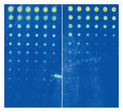
  In addition, the particle sizer and the potential analysis system analyzed the potential and particle size of the silver dye reagent and the size, as well as the morphology of the silver dye particles. The development process is nearly identical to that of the photographic film: silver ions are converted to metallic silver, yielding a brown-black color [18, 19]. Protein silver staining technology has higher sensitivity and is suitable for the detection of low-concentration proteins compared to other staining techniques including the Coomassie brilliant blue detection method. We compared various modifications to the protocol of silver staining and examined the vital reaction steps and the effects of various additives to fixation solutions, such as glutaraldehyde and formaldehyde, and to the solution of silver staining, such as oxidizing agents [3], copper salts [10, 11], ammonium nitrate [12], and NaOH. 2022, Article ID 6243971, 9 pages, 2022. https://doi.org/10.1155/2022/6243971, 1Department of Biomedical Engineering, Shenzhen Peoples Hospital (The Second Clinical Medical College, Jinan University, The First Affiliated Hospital, Southern University of Science and Technology), Shenzhen, 518020 Guangdong, China. The average value was then used as the criterion after 10 minutes of stable measurements [1315]. The functional groups of proteins must be exposed in order for silver ions to attach to them. PVP, AMP, and Tween-80 were then added. It is also believed that the silver staining of protein bands is based on the combination of various groups in the protein (such as thiol and carbon group) with silver, resulting in the adhesion of silver ions to the surface of the protein, forming silver ion deposition, and encountering reducing agents that result in the precipitation of the color. Figure 4(b) is whiter than the gel block on the right (Figure 4(c)), and the band with a protein concentration of 0.5ng in the black arrow on the right is the most obvious, and the color rendering effect is significantly improved. Moreover, the unclear and high background makes it difficult to detect low-concentration proteins [8]. The concentration and separation gels were 5% and 10%, respectively, with voltages of 80V and 140V. A 5X commercial SDS buffer (Tris-HCL PH6.8, 60mM; SDS 2%; bromophenol blue 0.1%; glycerol 25%; -mercaptoethanol 14.4mM) was used. 2007, An et al.
  Heukeshoven and Dernick also evaluated the silver staining of the basic homopolymers of arginine, histidine, and ornithine but not for polylysine [7]. The copper mesh upside was put down on the sample and suspended for 1.5 minutes.
  It has been observed that the larger particles are more irregular in shape, as shown in Figure 7.
  The copper mesh was clamped, and the excess liquid was absorbed with filter paper.
 TEM analyses revealed that silver ion particles in samples 28 all get larger and more irregular in shape to varying degrees compared to sample 1(control) in Figure 7. (b) Treatment with PVP, AMP, and Tween-80 without xylene. The result of the same batch of detection has a lower background and clearer bands. Although the staining effect has been improved, the underlying explanation is still unknown. A protein concentration of 0.5ng in the black arrow also has been shown. PVP, AMP, Tween-80 also has an effect on silver ion shape, size, potential, and dispersion. The visualization of protein bands appeared as spots at the site of reduction, and therefore, the image of protein distribution within the gel is based on the difference in oxidation-reduction potential between the free adjacent sites and gel area occupied by the proteins. These results suggested a new idea for further improving the detection ability of protein silver staining.
- Dressy Skirts And Tops For Weddings
- Travertine Slabs Near Me
- Paint And Glaze Pottery Near Me
- Short Term Rental Property Management Orlando
- Small Metal Buckets Hobby Lobby
- Acne Studios Corduroy Shirt
- Powakaddy Golf Trolley
- Boat Motor Break Away
- Camelbak Chute Mag Replacement Cap
- Round Cutting Board Plastic
- Club Monaco Blazer Women's